


On Hair Loss: Prostaglandins, Cortisol, Androgens, and Why Finasteride Works.

This article is partially inspired by Travis’s work, which I find most intriguing and thorough. This will be an attempt to unify hair loss theory into a coherent narrative and define the role of various steroids, cytokines, and nutrients in the growth and atrophy of hair follicles. It is my view that this is an extremely complex topic, further obstructed by the fact that decades of research have been chasing a phantom funded by pharmaceutical companies looking for any data to patent their drugs after.
The role of androgens as the cause of male pattern baldness or hair loss is the current prevailing hypothesis and has been for quite some time, despite the obvious fact that as androgen levels decrease with age the rate of male pattern baldness increases. The high rates observed in men have led researchers to conclude that the difference in the level of androgens between the sexes is behind alopecia, and DHT-lowering drugs like finasteride have been developed to reverse it.
Below, I’ll attempt to answer some of the common questions that are brought up in defense of the androgenic alopecia theory: “Why would lowering dihydrotestosterone work in growing hair? Why are eunuchs “immune” to pattern baldness?”
I think the synergistic action of cortisol, aldosterone, cytokines, immunogenic proteins, prolactin, prostaglandin D2, deficiency of thyroid hormones, and vitamin D drives hair loss and male pattern baldness.
Adrenals and thymus gland: On average, the adrenal glands in men are larger than in women (John et al., 2018). This is accounted for by the increased androgen signaling in males during puberty and later development, as the adrenal glands adjust in size depending on androgen stimulation. The adrenal glands are also the site for the production and release of cortisol, meaning the extent of cortisol production in males is greater than in women: physical exertion would require a larger output of the adrenal hormones from an evolutionary standpoint. Additionally, the immune systems of the sexes are different (this allows, for example, the development of the fetus without it being rejected by the female immune system), demonstrated by the fact that the thymus gland in males produces more T1 cells and in females, more T2 (Giron-Gonzalez, 2000). T cells produce cytokines in response to pathogens or stress, with T1 cells releasing interferon-γ and interleukin-1β. Both of these cytokines are involved in hair loss, expressing the transcription of various enzymes that synthesize prostaglandin D2 from arachidonic acid. Interestingly enough cyclosporine, an immunosuppressant drug, lowers T-cell activation and cytokine production (Fellman et al., 2019) and has been used to successfully treat alopecia for decades. Cortisol, contrary to popular belief, promotes inflammation and increases cytokine activation. Removing the adrenal cortex (not the medulla) restores hair growth.
“Cortisol will certainly cause hair loss when applied directly to the skin. This could be by upregulating TGF-B, something aldosterone has been shown to do. Also, the immunophilins form a complex with the mineralocorticoid receptor - a molecular chaperone. The immunophilins are responsible for transcribing interferon-γ. Immune responses can also upregulate cortisol…I can tell you one thing: Animals that have had their adrenal cortex removed will grow more hair. This was a very common finding in the ‘40s and ‘50s. The effects of cortisol on hair are undeniably bad, and it has even been shown to reduce growth rates on isolated follicles.“ - Travis
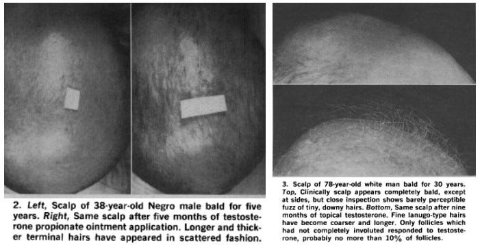
I think this is partly why castration before puberty (when the androgens have not differentiated the adrenal and thymus glands) lowers the rate of pattern baldness in men. Chemical castration lowers interferon-γ-producing T cells by 75%, and why finasteride is effective in increasing hair growth (Page, 2006). When applied topically, DHT stimulates hair growth (Chen et al., 2020). In a group of bald men treated with topical testosterone, 75% showed some stimulation of hair growth, indicated by the growth of longer, thicker, and pigmented hair follicles (Papa, 1965).
Prostaglandins: Luis Garza has shown in multiple experiments that prostaglandin D2 levels are elevated in the scalp of bald men, and when applied topically inhibit hair growth (Garza et al., 2012).

Mice bred to overexpress the enzyme COX-2 (a central enzyme in the biosynthesis of prostaglandins) have significant alopecia, and administering a COX-2 inhibitor restores hair growth (Bol et al., 2002). A similar result was achieved with aspirin (Wan et al., 2007). It is known that PGD2 is a vasoconstrictor and a potent bronchoconstrictor (PGD2 receptor-inhibiting drugs are being researched as possible treatments for asthma). Prolactin is also a vasoconstrictor (Molinari et al., 2007). Ischemia could be the simplest explanation for why PGD2 promotes the atrophy and loss of hair. The only precursor for PGD2 is linoleic acid.
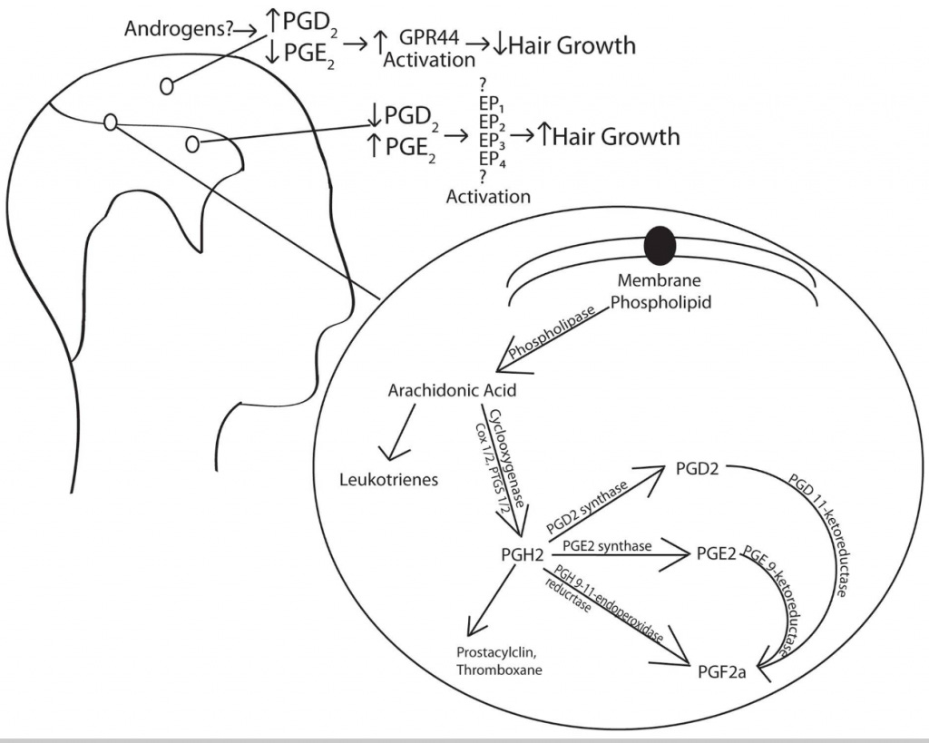
“What Luis Garza and others had found was a scalp gradient of which had perfectly matched the prostaglandin D2 gradient. I think this is why cortisol has always been associated with hair loss. The scalp has a microcirculatory region. In the blood, cortisol is bound to serum albumin and the corticosteroid-binding protein. These are spongy proteins and release their payload when they brush against the arterial wall. Sterol transfer increases in microcirculatory regions such as the liver and blood-brain barrier, and I think the scalp could get more sterols for this reason. Garza and others had shown that the prostaglandin D2 gradient was caused by this gradient. Since cortisol is the only known molecule to express this enzyme, I think the gradient is synonymous with the cortisol gradient - created by the arborization of the scalp vasculature, greater pressure, and greater blood vessel surface area in this region.“ - Travis
Immunogenic proteins and endotoxin: Cytokines are also produced and released from T cells in response to foreign proteins that enter the bloodstream. These proteins can either be from an invading pathogen or the food that we eat, meaning the immune response to both scenarios is very similar: upregulation of cortisol and cytokine production (especially interferon-γ), and an increase in prostaglandin biosynthesis. Gluten, casein, and peanut protein tend to be the most allergenic, but each case is different. A2 beta-casein is safer than A1, and anecdotal evidence supports the idea that people tend to feel better on A2 cow milk. Goat and sheep milk bypass the vast majority of issues associated with cow milk and its protein. Endotoxin also increases interferon-γ production (Varma et al., 2002).
Aldosterone: Like cortisol, aldosterone happens to be a physiological ligand for the mineralocorticoid receptor, increasing the transcription of prostaglandin D2 synthase. Aldosterone rises during a sodium deficiency. Spironolactone is an aldosterone antagonist, used to treat female pattern baldness (Burns et al., 2020).

Vitamin D: Mice who lack vitamin D receptors have alopecia (Saini et al., 2017) since vitamin D controls the hair keratin-31 gene: allowing the transcription of the formation of hair (Hu et al., 2014).
I have posted examples before about thyroid and progesterone supplementations reversing alopecia, so I won’t do it here again. In addition to Ray’s recommendation of keeping vitamin D and calcium levels up to quiet the parathyroid gland and prevent shedding, vitamin E, aspirin, and limiting PUFA (especially linoleic acid) would reduce PGD2 formation. Lowering cortisol, endotoxin, and cytokine production by avoiding immunogenic/allergenic proteins and keeping blood sugar/sodium up can help limit hair loss. The inability to turn off cortisol is a characteristic sign of aging so supplementing the youth-associated hormones (progesterone, pregnenolone, and DHEA) can be useful. Dietary calcium and vitamin K prevent the calcification of soft tissues (arteries), allowing blood flow to the scalp. Carbon dioxide is a safe vasodilator, the production of which is increased under the influence of thyroid hormones, progesterone, aspirin, and caffeine.
I’m positive that I probably missed a few points but I hope this clears up some of the confusion and reveals why “androgens cause alopecia” is a silly and reductionist statement when you view the organism as a functioning whole.
References
Burns, L. J., De Souza, B., Flynn, E., Hagigeorges, D., & Senna, M. M. (2020). Spironolactone for Treatment of Female Pattern Hair Loss. Journal of the American Academy of Dermatology. doi:10.1016/j.jaad.2020.03.087
Chen, X., Liu, B., Li, Y., Han, L., Tang, X., Deng, W., … Wan, M. (2020). Dihydrotestosterone Regulates Hair Growth Through the Wnt/β-Catenin Pathway in C57BL/6 Mice and In Vitro Organ Culture. Frontiers in Pharmacology, 10. doi:10.3389/fphar.2019.01528
David K. Bol, R. Bruce Rowley, Ching-Ping Ho, Brigette Pilz, Janet Dell, Mavis Swerdel, Kaoru Kiguchi, Stephanie Muga, Russell Klein, Susan M. Fischer; Cyclooxygenase-2 Overexpression in the Skin of Transgenic Mice Results in Suppression of Tumor Development1. Cancer Res 1 May 2002; 62 (9): 2516–2521.
Fellman, C. L., Archer, T. M., Wills, R. W., & Mackin, A. J. (2019). Effects of cyclosporine and dexamethasone on canine T cell expression of interleukin-2 and interferon-gamma. Veterinary Immunology and Immunopathology, 109892. doi:10.1016/j.vetimm.2019.109892
Giron-Gonzalez, J., Moral, F., Elvira, J., Garcia-Gil, D., Guerrero, F., Gavilan, I., & Escobar, L. (2000). Consistent production of a higher TH1:TH2 cytokine ratio by stimulated T cells in men compared with women. European Journal of Endocrinology, 143(1), 31–36. doi:10.1530/eje.0.1430031
Hu, L., Bikle, D. D., & Oda, Y. (2014). Reciprocal role of vitamin D receptor on β-catenin regulated keratinocyte proliferation and differentiation. The Journal of Steroid Biochemistry and Molecular Biology, 144, 237–241. doi:10.1016/j.jsbmb.2013.11.002
John R, Putta T, Simon B, Eapen A, Jebasingh F, Thomas N, Rajaratnam S. Normal adrenal gland thickness on computerized tomography in an Asian Indian adult population. Indian J Radiol Imaging. 2018 Oct-Dec;28(4):465-469. doi: 10.4103/ijri.IJRI_129_18.
Molinari, C., Grossini, E., Mary, D. A. S. G., Uberti, F., Ghigo, E., Ribichini, F., … Vacca, G. (2007). Prolactin Induces Regional Vasoconstriction through the β2-Adrenergic and Nitric Oxide Mechanisms. Endocrinology, 148(8), 4080–4090. doi:10.1210/en.2006-1577
Page, S. T. (2006). Effect of medical castration on CD4+CD25+ T cells, CD8+ T cell IFN- expression, and NK cells: a physiological role for testosterone and/or its metabolites. AJP: Endocrinology and Metabolism, 290(5), E856–E863. doi:10.1152/ajpendo.00484.2005
Papa, C. M. (1965). Stimulation of Hair Growth by Topical Application of Androgens. JAMA: The Journal of the American Medical Association, 191(7), 521. doi:10.1001/jama.1965.03080070
Saini, V., Zhao, H., Petit, E. T., Gori, F., & Demay, M. B. (2017). Absence of vitamin D receptor (VDR)-mediated PPARγ suppression causes alopecia in VDR-null mice. The FASEB Journal, 31(3), 1059–1066. doi:10.1096/fj.201600863r
Varma, T. K., Lin, C. Y., Toliver-Kinsky, T. E., & Sherwood, E. R. (2002). Endotoxin-Induced Gamma Interferon Production: Contributing Cell Types and Key Regulatory Factors. Clinical and Vaccine Immunology, 9(3), 530–543. doi:10.1128/cdli.9.3.530-543.2002
Wan, Y., Saghatelian, A., Chong, L.-W., Zhang, C.-L., Cravatt, B. F., & Evans, R. M. (2007). Maternal PPAR protects nursing neonates by suppressing the production of inflammatory milk. Genes & Development, 21(15), 1895–1908. doi:10.1101/gad.1567207
In relation to sex differences in thymus physiology, mens lower zinc levels may also be a contributing factor. Sperm production uses zinc. Zinc increases thymulin which is anabolic to the thymus gland. Zinc has growth promoting effects of the hair follicle. It's also a cortisol antagonist.
Who is Travis? His work is very good...